Quick Select:
Screening mammography changes breast cancer from a deadly disease to a manageable condition.
In line with international benchmarks, it is recommended that women over the age of 40 have a mammogram once a year. The screening interval is an important consideration. The longer the screening interval (i.e. the period of time between screening visits), the more cancers are discovered in the interval between screening visits and the less the benefit will be to the patient, as cancers could have been detected earlier.
Early detection by high-quality mammographic screening at specialised centres with stringent quality controls is the best method proven to decrease breast cancer mortality.

Ultrasound of the breast is not a primary screening tool but plays an important role in the further characterization of abnormalities identified at mammography.
The correct interpretation of a mammogram is crucial, and it is therefore essential that the practitioner is not only experienced in his/her field but remains at the forefront of scientific and technical advances in breast health.
High quality mammography is the single most efficient imaging modality for diagnosis of breast disease
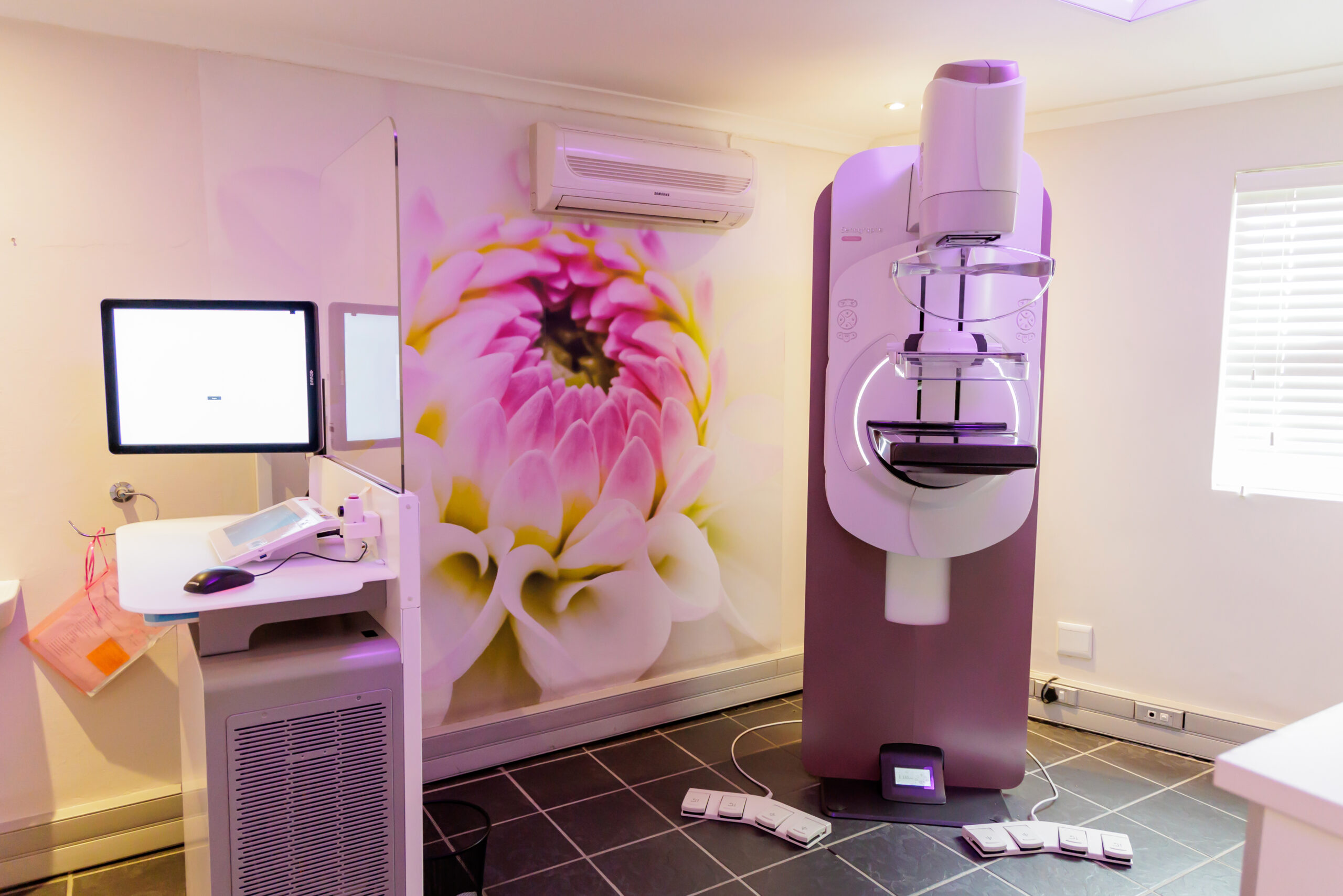
The new GE Seno Pristina, housed on-site, offers the best in Artificial Intelligence (AI) detection technology, using low compression techniques- allowing for enhanced detection and comfort.
Mammography is a highly specialised area of breast imaging. It is defined by the quality of equipment used, the technical skills required to perform the procedure correctly and the expertise of the professionals interpreting the mammogram.
A mammographer is a diagnostic radiographer who has an additional qualification in mammography and ultrasound of the breast. Maintaining excellent quality control is very important, resulting in earlier detection of breast cancer.
Several large studies conducted around the world have confirmed that regular screening mammograms can reduce the number of deaths from breast cancer for women aged between 40 to 74, especially for those over age 50, by more than half.
Early breast cancers can present in many ways on a mammogram. Although an experienced physician can detect a palpable mass, high-quality mammography is able to detect a non-palpable lesion up to 3 years before it becomes palpable.
A mammogram, when performed by an experienced mammographer, is not a painful procedure. At most, it may be a little uncomfortable. However, the short-term discomfort outweighs the long-term benefits.
Ultrasound imaging, also called ultrasound scanning or sonography
It is a non-invasive medical test using high-frequency sound waves to produce sonographic images. Ultrasound imaging does not use ionizing radiation (as used in X-rays).
Ultrasound of the breast is not a primary screening tool but has an important role in the further characterization of abnormalities identified at mammography and serves as a supplementary “double-check” to mammography.
High-definition breast ultrasound is therefore a very important and integral part of our protocol.
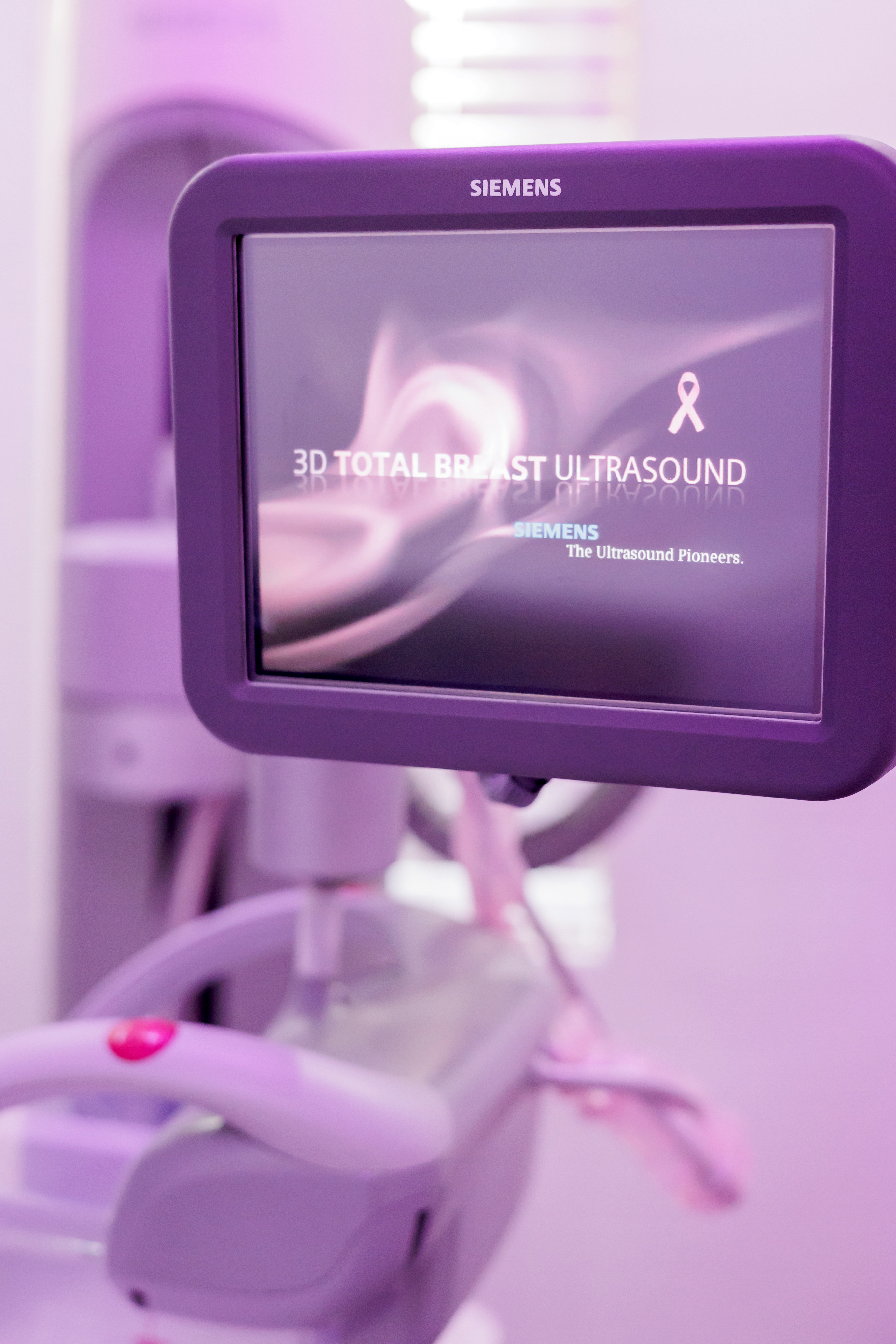
At our practice, a breast ultrasound is included, as a routine examination, with every 3D mammogram with AI-aided cancer detection, for every patient.

The most important uses of ultrasound in breast imaging are:
- As a primary screening tool for women under the age of 40, who do not yet require a mammogram.
Cyst vs. solid characterisation of clinically occult mammographically detected or palpable breast masses. - Evaluation of asymmetric tissue on mammography.
- Evaluation of palpable masses in women who are pregnant, lactating or under 40 years of age.
- Examination of the axilla for enlarged lymph nodes or other masses.
- Guide for interventional procedures (cyst aspiration, preoperative localization, fine needle aspiration and core biopsy).
Have a look at: FAQ’s | News & Events


